Empowering the clinician: putting pressure injury prevention at their fingertips
Arjo MOVE® — Provizio® SEM Scanner
Excecutive summary
The Provizio SEM Scanner is a FDA approved device that detects and measures localized edema or sub-epidermal moisture (SEM) earlier in the damage cycle before damage manifests on the surface — creating a critical window of opportunity for healthcare practitioners to provide the right interventions at the right anatomy that is developing pressure damage. There is a plethora of clinical evidence related to device functionality, clinical utility and standard of care published on the device.
The goal of this white paper is to analyze and summarize the quantitative findings from 11 healthcare facilities in the United States (US) who evaluated the Provizio SEM Scanner over recent years. Also presented are the qualitative results of feedback from clinical staff who used the device during the evaluation periods.
Introduction
Despite extensive research and resources employed for pressure injury (PI) prevention, hospital-acquired pressure injuries (HAPIs) remain a harmful and costly challenge to both patients and facilities.
The current standard of care for assessing a patient at risk of developing a PI has historically fallen short, especially for patients with dark tone skin. It is estimated that over 2.5 million acute care patients develop a PI1 every year in the US costing over 26.8 billion dollars.2 According to the Agency for Healthcare Research and Quality (AHRQ), HAPIs are the only recognized hospital acquired condition that has increased rather than decreased over recent years.3
Background
Current standard of care for assessing a patient’s risk for the development of a PI is a standardized and validated risk assessment tool (RAT) such as the Braden scale which is utilized extensively in the US. The participating facilities in this paper all utilized Braden as their RAT. Once the score is calculated, prevention interventions are initiated if the patient is scored to be at risk. A visual skin assessment (VSA) is usually completed at the same time to determine if there is an existing pressure injury noted. For anyone who has completed a root cause analysis (RCA) for a HAPI, they are acutely aware of the subjectivity noted in both the Braden scores documented and the VSA. Two clinical team members trained in risk assessment can review the same patient at the same time and based on their assessments, can obtain inconsistent Braden scores and possibly different VSA results. If the RAT or the VSA is the trigger for prevention to be initiated, this can explain why there may be a delay or omission of preventive interventions. Based on facility policy and national guidelines, these assessments are completed on patient admission to the facility and repeated, usually once a shift, dependent on patient condition. Both VSA and RATs are known to be subjective, and RATs are not anatomically specific and have a low predictive value.4,5
The color of the skin or possible lesion is interpreted by the assessing clinician. In dark skin tones, non-blanchable erythema may be difficult to distinguish from surrounding tissue, hence a Stage 1 PI could be missed, and subsequently any timely intervention. Patients with dark skin tones are four times more likely to die from PI related causes,6 have a 60% greater risk of PIs than light skin tone patients,7 have a greater risk of multiple PIs and suffering worst stage PIs.8
The 2019 NPIAP International Clinical Practice Guidelines recognizes sub-epidermal moisture assessment as an important factor in PI prevention practice. Recommendation 2.6 states “Consider using a sub-epidermal moisture/edema measurement device as an adjunct to routine clinical skin assessment” and recommendation 2.7 states “When assessing darkly pigmented skin, consider assessment of skin temperature and sub-epidermal moisture as important adjunct assessment strategies.”9
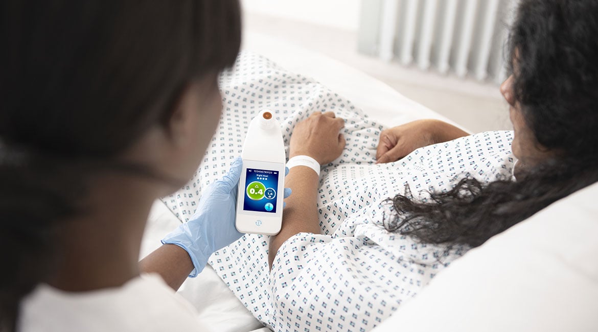
The Provizio SEM Scanner is a non-invasive device that is used for detection of sub-epidermal moisture before it is visible on the skin, by measuring fluctuations in SEM, also known as localized edema and persistent focal edema. It is a “point of care” device to provide rapid results that compares SEM measurements across an anatomical site and reports the difference between values at the inflamed versus healthy tissue site — this is the SEM Delta (Δ).10 A Delta that is ≥0.6 should trigger early intervention and subsequently decrease HAPI by treating the raised levels of SEM.11 The heels and sacrum are the anatomical sites that have been approved for assessment by the scanner, and these areas are most commonly where PIs develop.12
Arjo partnered with eleven facilities for evaluation of the device. The facilities ranged from large academic teaching institutes and trauma centers to community hospitals. Each of these facilities chose one or more patient care units to utilize the Provizio SEM Scanner as part of their daily assessment as an adjunct to their usual PI risk assessment protocol. The evaluation period for each facility lasted one to four weeks.
Methodology
The methodology employed was to have the bedside staff nurse, wound care nurse or designated unit RN scan the heels and sacrum of the patient upon admission to the unit and during one or both of the daily assessments. The SEM Deltas (Δ) were recorded and compared to the Braden scores and VSA over the same time period. Interventions were implemented based on the SEM Delta (Δ) readings for the specific anatomical site. The data was then reviewed to determine if the Provizio SEM Scanner indicated patients at risk of a PI that were not identified by the Braden score or VSA.
Implementation
All staff involved in the evaluation process were trained by an Arjo MOVE Clinical Consultant in the “damage cascade.”14 This explains the ”why” of how the scanner detects SEM before damage is visible on the skin, despite a normal skin assessment and perhaps a higher Braden score that does not indicate that PI prevention is needed. The nurse is also trained in the use of scanner and where the scanning is performed on the heels and sacrum and its role in detection and measurement of localized edema or SEM earlier in the damage cascade before damage manifests on the surface. Their own facility policy on PI prevention interventions is then reinforced so the bedside nurse knows when to initiate the targeted and early interventions before the tissue damage is visible on the skin. In addition, the facility staff are provided tips on how to integrate the scanning into their routine “head-to-toe” assessment. On site intensive follow up is provided by the Arjo MOVE Clinical Consultant during the evaluation process to assist in reinforcing scanning compliance and timely clinical interventions based on an elevated SEM Delta (Δ).
In order for a new technology or process to be implemented into a healthcare provider’s standard of care, it needs to be embraced and advocated by senior leadership, in addition to the staff at the bedside utilizing the technology. An article written by Zach Smith, BSN, RN, VP of Brand at Stability Healthcare, lists 3 reasons why new technology fails when released into a hospital:
- It is too difficult to learn
- It does not resonate with the staff
- It doesn’t solve an existing pain point with the user13
The Provizio SEM Scanner answers all of the above aspects. It is not a complex device to learn how to operate, it empowers all levels of clinical staff and it helps identify patients at risk of developing a HAPI, which is a quality metric that all members of the multi-disciplinary team are concerned about.
Results
Although there were many metrics collected, this paper will focus on the identification of at-risk patients by the Provizio SEM Scanner and interventions initiated that may have been missed based on RAT and VSA alone. In total, 689 patients were scanned over the course of the eleven evaluations with 4,297 scans completed. Results are divided into 2 categories, quantitative and qualitative.
Quantitative results
Analysis of the data revealed that the Provizio SEM Scanner noticeably increased the user’s ability to identify which patients were at increased risk of developing a PI at an anatomically specific area and had clinical interventions initiated to alleviate PI risk. Without the scanning, these “at-risk” patients may not have received clinical interventions, and subsequently could have gone on to develop a pressure injury.
- 227 patients (33%) were identified as having subcutaneous tissue damage by the Provizio SEM Scanner but had no skin discoloration and a RAT score that indicated ”low risk” (Braden score 15-18) or “not at risk” (Braden 19-23) for PI development
- 43% of clinical decisions were made based on the Provizio SEM Scanner Delta (Δ)
- 58% of clinical decisions included increased turning or mobilization
- 11% of clinical decisions included the introduction of a new support surface. This number ranged from 0 to 50% in the facilities as many of the evaluations were completed in an intensive care unit where they already had higher level support surfaces
- 43% of patients had heel support and/or heel elevation initiated based on the Provizio SEM Scanner Delta (Δ)
Qualitative results
Five of the eleven facilities completed a short survey post evaluation to gather feedback from the nurses who performed the scanning. Thirty-six nurses completed the survey which included questions related to the ease of use, how well the device increased their ability to identify patients at increased risk of developing a PI and whether the device increased their ability to understand whether the clinical interventions applied were effective at reducing the risk of a PI developing. The results of the surveys are summarized as follows.
- 100% of nurses responded that it was “extremely or somewhat easy” to use the Provizio SEM Scanner
- 94% found the Provizio SEM Scanner to be a useful tool for anatomy specific patient assessment
- 92% felt that the training increased their ability to identify patients with increased risk of a pressure injury
- 100% indicated that they were extremely satisfied or satisfied with the training of the Provizio SEM Scanner
- 94% felt that the training increased their ability to understand whether the clinical interventions they applied with patients at increased risk of pressure injury were effective
Some specific comments from nursing staff that completed the survey included:
- “Training increased my ability to know whether the clinical interventions I applied with patients at increased risk of pressure injury were effective.”
- “HAPIs can be prevented using the scanner. Nurses/PCA applied preventative measure when readings were high.”
- “A great addition and resource to help this vulnerable patient population.”
- “The more technology we have to take care of patients, the better it is to deliver quality care. Great tool!”
Conclusion
Hospital acquired pressure injuries continue to be a challenge to both facilities and the patients affected by this problem. Despite advances in prevention products, training and risk assessment processes implemented by hospitals, patients continue to develop PIs. Based on standard of care, patients are assigned a PI risk score in addition to a visual skin assessment that should identify those at risk, and when prevention strategies should be employed. The current standard of care is not adequate and HAPIs develop. The implementation of the Provizio SEM Scanner objectively identifies at risk individuals that may have been missed on routine assessments especially in patients with dark skin tone. This allows for anatomically specific interventions to be initiated earlier, before the damage is visualized on the skin. The Provizio SEM Scanner places objective, early and targeted risk identification to prevent HAPIs in the hands of the caregiver.
Click here to download the white paper.
The Science of Sub-Epidermal Moisture (SEM) clinical evidence summary
Elevated levels of SEM is a biomarker of early tissue damage that can lead to pressure injury development. SEM can be identified by assessing the biocapacitance of tissue. This noninvasive technology enables early and objective assessment of increased pressure injury (PI) risk, empowering you to take decisive action to minimize PI incidence and to help reduce overall cost and time to care.
Download our Science of SEM clinical evidence summary and learn about:
- The challenges of preventing pressure injuries
- Effects of prolonged pressure on tissue
- The Provizio SEM Scanner hand-held wireless, noninvasive device
- Foundational clinical studies
DOWNLOAD SCIENCE OF SEM CLINICAL EVIDENCE SUMMARY
Talk to an Arjo Expert
Learn more about the Provizio SEM Scanner by speaking with an Arjo Expert, who will respond to your request in a timely manner.
References:
- Berlowitz D, VanDeusen Lukas C, Parker V, et al. (Content last reviewed October 2014.) Preventing Pressure Ulcers in Hospitals: A Toolkit for Improving Quality of Care. Agency for Healthcare Research and Quality, Rockville, MD. Retrieved from http://www.ahrq.gov/professionals/systems/hospital/pressureulcertoolkit/index.html.
- Padula WV, Delarmente BA. The national cost of hospital-acquired pressure injuries in the United States. Int Wound J. 2019 Jun;16(3):634-640. doi: 10.1111/iwj.13071. Epub 2019 Jan 28. PMID: 30693644; PMCID: PMC7948545.
- AHRQ National Scorecard on Hospital-Acquired Conditions. Content last reviewed July 2020. Agency for Healthcare Research and Quality, Rockville, MD https://www. ahrq.gov/hai/pfp/index.html (Link).
- Moore ZEH, Patton D. Risk assessment tools for the prevention of pressure ulcers. Cochrane Database of Systematic Reviews 2019, Issue 1. Art No.:CD006471. DOI:10.1002/14651858.CD006471.Pub4.
- Samuriwo R. & Dowding D (2014) Nurses’ pressure ulcer related judgements and decisions in clinical practice: A systematic review. Int J Nurs. 51(12):1667-85.
- Redelings, Matthew D et al. “Pressure ulcers: more lethal than we thought?” Advances in skin & wound care vol. 18.7 (2005): 367-72. doi: 10.1097/00129334- 200509000-00010.
- Baumgarten, Mona et al. “Black/White differences in pressure ulcer incidence in nursing home residents.” Journal of the American Geriatrics Society vol. 52.8 (2004): 1293-8. doi: 10.1111.j.1532-5415- 2004.52358.x.
- Gunowa, N et al. “Pressure Injuries in People with Dark Skin tones: A Literature Review.” Journal of Clinical Nursing, vol. 27, 2018, pp. 3266-75.
- European Pressure Ulcer Advisory Panel, National Pressure Injury Advisory Panel and Pan Pacific Pressure Injury Alliance. Prevention and Treatment of Pressure Ulcers/ Injuries: Quick Reference Guide. Emily Haesler (Ed.). EPUAP/NPIAP/PPPIA: 2019.
- Gershon, S. et al, Journal of Wound Care, Vol. 30, No. 1, 2021.
- Rose Raizman, Minette MacNeil, Laurie Rappl. Utility of a sensor-based technology to assist in the prevention of pressure ulcers: A clinical comparison, Int Wound J. 2018 Dec;15(6): 1033-1044.
- Moore Z. et al. (2019). The prevalence of pressure ulcers in Europe, what does the European data tell us: a systematic review. Journal of Wound Care. 28(11) 710-719 (Link).
- Smith, Zach. The Key to Technology Adoption on Hospital Departments. (https://nursegrid.com/author/zach).
- Gefen, A., et al. (2020). Update to device-related pressure ulcers: SECURE prevention. COVID-19, face masks and skin damage. (Journal of Wound Care Vol 29, NO 5, May 2020.